1. Reading Chest X-rays With Reason, Not Just Answers
If you have spent time in a reading room, you know the rhythm. Scroll, zoom, compare priors, dictate, repeat. Most AI in medical imaging tools promise to make that loop faster. Fewer promise to make it safer. The difference comes down to one capability, showing the why behind a prediction, not only the what. This year, a new class of medical foundation models started doing both. They answer questions, and they show their work, grounded to the pixels that persuaded them. That sounds small. In practice, it is the shift that unlocks trust.
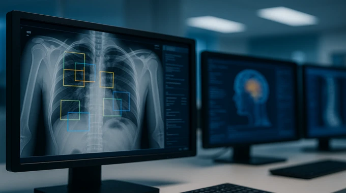
The headline case is a chest X-ray foundation model that couples free text reporting, visual question answering, and grounded reasoning. It is trained in stages, instruction tuning, synthetic step-by-step rationales to cold start reasoning, then online reinforcement learning to polish both the final answers and the intermediate steps. The outcome is a model that points to the right ribs, lines, and bases while it explains itself, rather than leaving clinicians to guess.
Table of Contents
2. What Changed In 2025, Grounded Reasoning Arrives
The model’s training recipe is refreshingly practical. Start with a strong general vision-language base, Qwen2.5-VL-7B, then instruction-tune it on hundreds of thousands of curated chest X-ray samples to get basic competency. Add a small but high quality set of synthetic reasoning traces that include bounding boxes, teach the model how to talk through a case and point to evidence. Finish with online reinforcement learning that scores both the answer and the reasoning path, and update the policy while keeping it close to a safe reference. The end result, DeepMedix-R1, consistently outputs an answer plus region-tied reasoning for each query.
Performance is the second change. On report generation, it improves over prior open models, with average gains versus LLaVA-Rad and MedGemma, and on VQA it clears CheXagent by a healthy margin. Even more interesting, human experts prefer its reasoning over a popular baseline by about three to one, which speaks directly to clinical plausibility.
3. How The Model Thinks, From Instructions To Online RL
Under the hood, the online phase uses a group relative policy optimization setup. For each question, the system samples multiple candidate outputs, then scores them along three axes. First, how close is the answer to ground truth, with exact match for closed answers, F1 for multi-label, and a blend of BLEU and ROUGE for free text.
Second, does the reasoning include valid image coordinates, and do those boxes stay inside the image bounds. Third, did the model follow the required output format. The relative rewards are standardized within a group, then used to update the policy, with a KL term that pulls the new policy back toward a safe reference. This keeps learning stable, and it encourages grounded, properly formatted, clinically useful steps.
Evaluation is equally thoughtful. Alongside conventional metrics, the authors introduce an LLM-as-judge setup, Report Arena, that compares paired reports and computes Bradley-Terry ranking scores. This gives a second, human-like lens on quality, and it mirrors how clinicians compare two reads.
4. Does It Actually Work, Benchmarks And Preference
Benchmarks cover four datasets, MIMIC-CXR and Open-I for report generation, Ext-VQA and CXR-VQA for question answering. DeepMedix-R1 tops prior medical and CXR-specific models on average, and the online reinforcement learning stage adds measurable lift across all splits. In plain language, the model writes better reports and answers more questions correctly, while also explaining itself better. That is the trifecta many teams have chased.
Expert review is the real test. Radiology annotators scored reasoning for relevance, correctness, completeness, and groundedness. The grounded reasoning preference and overall preference lean strongly toward the new system compared to a widely used baseline. That preference is what you want in a clinical deployment, because it correlates with whether a clinician will trust and adopt the tool in the first place.
5. Collaboration At The Edge, Agentic Triage That Knows When To Ask For Help

Speed is not the only goal. A good system must also know when to defer. Recent work on agentic triage for chest X-rays, AT-CXR, takes exactly that stance. It estimates per-case uncertainty and distributional fit, then routes studies through a simple policy. If confidence is high, the agent acts. If not, it abstains with a suggested label and hands off to a radiologist. The team reports stronger selective prediction, lower area under risk-coverage curves, and operating latency that fits clinical constraints. They also compare a rule based router with an LLM router, which gives deployment tradeoffs, throughput versus peak accuracy. That is the right design for real wards, where missing a tension pneumothorax is not an option.
6. Explainability That Clinicians Can Use
Grounded rationales are not the only way to make models legible. The broader body of explainable AI in healthcare offers a tidy taxonomy. You have attribution methods like Grad-CAM and SmoothGrad that highlight influential regions. You have perturbation methods that probe the effect of masking or noising patches. With modern transformers, attention maps add another handle. The tradeoffs are familiar, attribution is fast and easy to plug in, perturbation can be more faithful but slower, and attention must be interpreted with care. For medical imaging, the principle is simple. Explanations must be clinically meaningful, not only visually aligned boxes. A saliency blob with high overlap that points to the wrong side of the chest is a miss, not a success.
If your team builds detection style pipelines, tie the explanation back to the detection task. Boxes, labels, and confidence should line up with the narrative in the report. Do not ship a heatmap without a sentence that explains what that heatmap implies. Clinicians do not read pixels, they read evidence.
7. What This Means For AI Medical Imaging Companies

If you are an AI medical imaging company, the bar moved. Buyers will still ask for accuracy and throughput. Increasingly, they will also ask what the model saw, and why it made a call. The chest X-ray foundation model above shows a pattern you can reuse. Build an instruction tuned base over medical data, add a small but clean set of grounded reasoning traces, then use online reinforcement learning to shape both the final answer and the intermediate steps. This is a practical path to explainable AI in healthcare that does not sacrifice performance.
Operationally, invest in three things. First, an evaluation bed that blends text metrics, fact metrics like RadGraph or CheXbert, and pairwise human or LLM-as-judge comparisons. Second, a selective prediction stack that can abstain intelligently and route cases. Third, a policy for how explanations show up in the reading workflow, bounding boxes, structured rationale, hyperlinks from text spans to regions, and audit trails.
8. Quick Comparison, When To Reach For What
| Approach | Best For | What You Get | Typical Failure Mode | Integration Tips |
|---|---|---|---|---|
| Medical foundation model with grounded reasoning, for example DeepMedix-R1 | Report generation, VQA, teaching settings | Free-text output with stepwise rationale tied to image regions | Fluent text that hides a wrong step if grounding is weak | Render rationales inline, link each reasoning step to a visible box, log both answer and steps for audit. |
| Agentic triage, for example AT-CXR | High volume front-doors, urgent care triage | Selective prediction, abstain and escalate under time budgets | Over- or under-abstaining on distribution shift | Tune the router to site constraints, decide if rules or an LLM router fit your latency and governance requirements. |
| Classic XAI add-ons, attribution and perturbation | Quick audits, model debugging, education | Heatmaps and masks that suggest important regions | Pretty plots without clinical meaning | Pair every map with a one-line interpretation. Use consistent color scales. Prefer task linked overlays. |
9. A Simple Build Plan For Hospitals And Vendors
9.1 Collect The Right Supervision
You do not need millions of grounded rationales. You do need a few thousand that are high quality. Focus on common findings, pleural effusions, cardiomegaly, atelectasis, lines and tubes, and common error modes where grounding helps. The chest X-ray model above reached useful reasoning quality with thousands of curated reasoning traces and a larger instruction set. That is achievable for many teams.
9.2 Train With Guardrails
Keep a reference model and use a KL pull during online learning so your policy does not drift. Reward both answers and coordinates, and penalize coordinates that wander off image bounds. Enforce output formats that your UI can render deterministically. These small details compound into reliability.
9.3 Evaluate Like A Clinic, Not A Kaggle
Blend text similarity, fact extraction, and paired comparisons. Measure coverage risk for selective prediction. Track preference by radiologists for the reasoning itself, not only final answers. The biggest wins show up when both the answer and the path are good. That is what clinicians trust.
9.4 Design For The Reading Room
Do not bury explanations in a separate tab. Tie them to the viewport. When a sentence mentions a blunted costophrenic angle, a click should jump to that corner. When the agent abstains, it should say what finding is uncertain and why. When the model is confident, it should still show what evidence made it confident. That is how you turn explainable AI in healthcare from a checkbox into a daily advantage.
10. Where AI In Radiology Goes Next
AI in medical imaging is converging on a sensible middle ground. On one side, general models that can read, localize, and explain across tasks. On the other side, agentic systems that know when to push and when to pass the case to a human. Both are wrapped in explanation layers that respect clinical context. The chest X-ray foundation model shows that you do not have to trade accuracy for legibility, and triage agents show that abstention is a feature, not a flaw. Together, they push the future of AI in medical imaging toward collaboration, not replacement.
If you build products, push past glossy demos. Show grounded rationales that match anatomy. Prove selective prediction under your site’s timelines. Publish a clear evaluation story with both metrics and pairwise comparisons. If you run a department, pilot tools that can explain themselves, and insist that abstain and escalate are first class behaviors. The result is simple. Better reports, safer workflows, and clinicians who trust what the model says because they can see how it got there.
Call to action. If you lead an AI medical imaging company, ship one workflow this quarter that links every finding in a report to the exact pixels that justify it. If you lead a hospital program, pick one high volume pathway, add an agentic triage that knows when to ask for help, and measure risk-coverage before and after. Then share the numbers. That is how we move AI in medical imaging from promise to practice.
Notes And Sources
Key details on grounded reasoning, staged training, Report Arena, and expert preference are from the DeepMedix-R1 paper on chest X-ray interpretation.
Agentic triage summary and metrics language draw on the AT-CXR preprint description. (Cool Papers)
Background taxonomy for explainable AI in healthcare and computer vision references a recent comprehensive review. (MDPI)
How is AI used in medical imaging?
AI in medical imaging supports the end to end workflow. It helps with image acquisition and reconstruction, flags urgent studies for triage, detects and measures lesions, segments organs, and compares priors. It can draft structured or free text reports, suggest impressions, and run quality checks. In practice, these systems plug into PACS, RIS, and EHR so cases route to the right reader and findings stay consistent. Teams also use AI for dose reduction and faster scans, which improves patient comfort and throughput without sacrificing diagnostic quality.
What are the benefits of AI in medical imaging?
When deployed well, AI in medical imaging speeds interpretation, reduces variability, and improves sensitivity for time critical findings. Triage can move stroke or pneumothorax cases to the front of the queue. Automated measurements, segmentation, and report drafting save minutes per study, which adds up across a list. Dose and scan time reductions can lower risk and improve experience. The biggest gains come from good validation, clear operating points, and training plans, not from the model alone. Hospitals see value when AI aligns with real clinical workflow.
What are the challenges of using AI in medical imaging?
Key challenges include data quality, bias, and generalization across vendors, scanners, and sites. AI in medical imaging must integrate with PACS, RIS, and EHR, which needs IT time and change management. Explainability and uncertainty estimates are important for trust. Monitoring and drift detection keep performance stable after go live. Privacy, cybersecurity, and regulatory documentation add overhead. Success depends on governance, audit trails, and escalation rules. Many teams run a pilot with measurable endpoints, then scale only after the service level and safety metrics look strong.
Is AI going to replace radiologists?
No. AI in medical imaging is built to augment radiologists. Algorithms excel at repetitive pattern recognition, rapid triage, and measurement tasks. Clinicians lead on differential diagnosis, rare presentations, patient communication, and accountability. The role is shifting toward orchestrating AI assisted workflows, designing protocols, and reviewing edge cases. With good tools, radiologists spend less time on clicks and more time on complex decisions and multidisciplinary care. Human in the loop review remains essential for safety, quality, and legal responsibility.
What is a medical foundation model and how does it work?
A medical foundation model is a large model pretrained on diverse imaging data, often paired with reports. In AI in medical imaging, the model learns general visual and clinical patterns, then adapts to tasks like classification, segmentation, visual question answering, or report generation. Fine tuning uses curated labels or instructions. The strongest systems add grounded reasoning, which links each conclusion to image regions, plus uncertainty estimates that guide when to automate or escalate. Hospitals still need local validation, monitoring, and clear operating procedures before routine use.